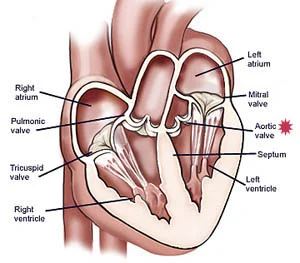
Between the four chambers that make up the human heart and the two large vessels coming out of the heart, there are valves that regulate blood flow and ensure that there is flow in one direction. During circulation, blood first comes to the right atrium of the heart. After passing through the tricuspid valve into the right ventricle, it travels through the pulmonary valve to the pulmonary artery and lungs.
When the blood reaches the lungs, it leaves the carbon dioxide and takes the oxygen and goes to the left atrium of the heart. After passing through the mitral valve to the left ventricle, the circulation cycle is completed by pumping it through the aorta to the aorta and the whole body.
The valve on the left side of the heart that regulates blood flow between the left ventricle and the aorta is called the aortic valve. Aortic stenosis due to calcification of the aortic valve is called aortic stenosis, and weakening of the valve tissue despite the absence of calcification is called aortic insufficiency.
Diagnosis of Aortic Valve Disease
In addition to general examination, echocardiography is used to diagnose aortic valve disease. With echocardiography, the internal structure and functionality of the heart are visualized, as well as data on heart functions, the size of the heart, and the degree of contraction. Depending on the evaluation of the patient’s complaints and echocardiographic examination results, a decision is made as to whether there is a need for intervention on the valves.
Causes of Aortic Valve Disease
Acquired aortic stenosis can be seen in the 20s and 30s due to rheumatic fever in childhood, or in later ages due to aging.
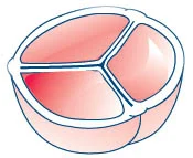
It may occur if the aortic valve, which normally consists of 3 leaflets, consists of two leaflets or a single leaflet. Since a single leaflet is not compatible with life, it necessitates a surgical procedure at birth.
In the formation of two leaflets, surgery may be required in childhood, as well as the need for surgery in case of stenosis due to calcification in the valve with advancing age. People with two-leaf aortic valves are also more likely to have an aortic aneurysm.
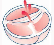
Aortic valve regurgitation occurs when the aortic valve does not close completely. In this case, some of the blood pumped by the heart goes back to the left ventricle of the heart. Fatigue and shortness of breath occur due to the inability of the heart to pump blood to the body fully.
Aortic valve insufficiency may occur due to:
- Congenital anomalies;
- Connective tissue diseases;
- Chest trauma;
- Rheumatic fever in childhood;
- Changes in the valve due to aging.
Aortic Valve Disease Treatment Methods
Aortic valve disease can be treated with valve repair and valve replacement. There are different methods that can be applied in valve repair and valve replacement procedures applied for this purpose.
Transcatheter Valve Treatment: In this method, which is an interventional procedure, valve repair or replacement can be performed by using clips or catheters through large blood vessels.
In cases where there is a leak in the mitral valve, backflow of blood can be prevented by attaching a latch at the ends of the valves within the scope of the procedure called Mitra Clip. The recovery period of the patients to whom this method is applied is shorter than the surgical procedure.
TAVI: Transcatheter Aortic Valve Implantation procedure can be defined as the current valve application for aortic valves recently. It is entered through the patient’s inguinal vein through the catheter. The procedure is completed by advancing a catheter with a balloon in the middle and an artificial valve folded around it to the level of the narrowed aortic valve, inflating the balloon at the valve level, opening the valve around it, and placing it inside the old valve.
The balloon is then deflated and withdrawn with the catheter. Thus, the new valve is put in place. The procedure, which is applied in order to be used in patients in the advanced age group and with high surgical risk, is being applied with an increasing frequency in patients in the middle risk group as a result of developments in technology and medicine.
The point to be considered in the treatment of aortic valve disease is to perform the surgery at the appropriate time. The determining factors in the surgery are the patient’s current complaints, aortic insufficiency or stenosis reaching critical levels. In cases where a surgical procedure is not required for the treatment of the patient, the patient is followed up regularly. Medication may be taken if deemed necessary.
Aortic Valve Repair
It is a method applied by placing the tissues that support the valve structure, providing better closure by cutting and repairing the valve tissue, or releasing the adhered leaflets.
However, valve repair is not always the case. Although valve repair is a more common method for mitral valves, aortic and pulmonary valves need to be replaced frequently.
It is possible to use a method called Balloon Valvuloplasty in the treatment of narrowed aortic valves. During the procedure, as in the angiography procedure, a catheter with a balloon at the tip is advanced to the level of the narrowed valve and the balloon is inflated to expand the narrowed valve. It is a method that provides effective results especially in the mitral valve.
Aortic Valve Replacement
One of the most frequently applied methods in the diseases of replacement of valve is valve replacement surgery. After the valve to be treated during the surgery is removed, a mechanical or biological valve is placed in its place.
Biological valves are obtained from pig, bovine or human tissue. In case of valve replacement using a biological valve, the use of blood thinners is not required, but it may be necessary to repeat the operation process within 10 to 15 years due to the wear of the biological valves. Therefore, it is not a preferred method in young patients.
Although mechanical valves can be used for a lifetime, it is a method that requires regular taking of blood thinners. If blood thinners are not taken, problems such as clogging of mechanical valves with clots or paralysis may occur.
In cases where aortic valve replacement is required, the use of a heart valve formed from the patient’s own heart membrane may also be an alternative. Patients undergoing this method, known as the Ozaki procedure, do not need to take anticoagulants. However, the problem of coagulation on the valve, which is at risk of being encountered in other prosthetic valves, is not usually seen in patients with this technique.
Considerations After Aortic Valve Surgery
It is temporary for the patient to be angry, depressed or nervous after aortic valve surgery. Some changes should be made in lifestyle habits in order to experience a faster recovery process after the surgery and to avoid any health problems.
Loss of appetite can be seen in the first weeks after the surgery. In the first month, care should be taken to feed frequently with small portions. In addition to applying a nutrition program suitable for heart health, plenty of water should be consumed.
The movements of the patient should be controlled in the first 2 months following the surgery. Otherwise, as a result of wrong movements, undesirable situations such as delay or prevention of union of the cut sternum during surgery may occur.
Reaching for objects above shoulder level should be avoided during the post-operative recovery period. During bending, care should be taken to bend from the knees, not from the waist, and to use the leg muscles instead of the arms when getting up from the sitting position.
In the first 2 weeks after the operation, stairs can be climbed, namely 1 floor per day, and then by gradually increasing. Care should be taken not to lift more than 5 kilograms for at least 1 month.
Prescribed drugs should be taken regularly and the chest corset should be used for 2 months. Especially when coughing and sneezing, the hands and the front of the corset should be held. In addition, embolism prevention stockings should be worn constantly.